Call us
 Email us
treatment of retina detachment
This depends on if you have developed a retina hole or tear or has it progressed to a retinal detachment.
Treatment for Retina Hole or Retina Tear
The hole or tear is sealed by producing minute scars on the retina. The scar tissue helps to “weld” the retina to the underlying support tissue and prevent fluid from seeping through the hole. This can reduce or stop the risk of progression to retinal detachment. These scars can be produced by:
-
Laser photocoagulation
using a laser or strong light source which is focussed throught the pupil on the retina.
-
Cryotherapy
controlled freezing of the affected retina tissue by using a frozen probe. Numbing eye drops are used to ensure minimal discomfort.
These are outpatient procedures and you do not need to get admitted for them.

Treatment for Retinal Detachment
If the retina has become detached and the detachment is too large for laser treatment or cryotherapy alone, surgery is necessary to “re-attach” the retina. Without some type of retinal re-attachment surgery, vision will almost always be lost.
Surgery for retinal detachment can be the following:
-
Scleral Buckling
Scleral buckling involves the sewing a piece of silicone to the white part (sclera) over the affected retina.
Scleral buckling is performed in the operating room under local or general anaesthesia.
It is an “extraocular procedure” – meaning the surgeon does not enter the eyeball like in a vitrectomy. In this process, a piece of silicone plastic is sewn onto the outside wall of the eye (sclera) over the site of the tear.These buckles and bands are left permanently and are not visible from outside.
Success rates for reattaching the retina with scleral buckling are approximately 90-95%.
-
Pneumoretinopexy
A gas bubble is injected into the vitreous cavity of the eye.
The gas bubble pushes against the outer wall of the eye and helps seal off fluid leaking into the vitreous through the gap in the retina.
The fluid that collected under the retina gets reabsorbed by the body. The retina can now stick to the eye wall like it should. Eventually, the gas bubble also gets reabsorbed.
This is why the patient is instructed to keep their head in a specific position so that the gas bubble seals the retina tear by its surface tension effect.
-
Vitrectomy
Vitreous surgery is now routinely undertaken for primary detachments. It is especially useful when:
The retinal tears are very large
The Tear is placed very far back (posteriorly) on the retina
A macular hole is causing detachment
If there is blood in the vitreous blocking a clear view of the retina
If there is proliferative vitreo-retinopathy
The vitreous is removed and replaced by air or oil. If silicone oil has been used, it has to be removed at a later date as a separate surgical procedure.
-
Microincision Vitreous Surgery (MIVS)
with smaller instrument size is the latest revolution in Vitreous surgery. The finer instrumentation allows one to perform technically complex steps with greater safety. It also allows us to safely operate closer to the retina for peeling of fine membranes from the surface of the retina.
Video Blogs
Watch & Learn


Our Retina Experts
Dr. Cyrus Shroff
Dr. Daraius Shroff
Dr. Sharad Rohatgi
Dr. Charu Gupta
Dr. Shishir Narain
Dr. Neelam Atri
Dr. Gagan Bhatia
Dr. Arindam Chakravarti
Dr. Stuti Astir
Dr. Priyanka Gupta
Dr. Sandeep Kumar
Dr. Minal Sharma
Dr. Aashraya Prithviraj Karpe
Dr. Richa Pyare
Dr. Minal Kaur
FAQs
-
What is the outlook for people with retinal detachment?
Your outlook or prognosis depends on factors like how your vision was before the retinal detachment, how extensive your detachment was and if there are any other complicating factors. Our advanced retina detachment surgery experts will always discuss this with you prior to surgery.
In general, surgery for retinal detachment is very successful — the repair works in a majority of the cases. Sometimes, you may need more than one procedure to return the retina to its place.
that just successfully reattaching the retina (anatomical success) does not always translate into marked visual improvement (functional success).
This is because fine vision may be permanently affected if the macula is involved; There is less visual return when the retina has been detached for a long duration, or there is a fibrous growth on the surface of the retina. This is why it is important to treat retinal detachment early.
-
What can you expect after retinal detachment surgery?
Retina surgery can be done under local or general anesthesia.We will keep you under observation for a few hours after surgery.
You’ll also need to take it easy for a few weeks. Check with our retina surgeons about when you can exercise, drive and get back to your regular activities.
Discomfort: After retinal detachment surgery, you may have some discomfort. It can last for a few weeks. We will prescribe basic pain medicine to help you during this time.
Take it slow & easy: We may ask you to avoid sudden jerks, driving and exercise for a few weeks.
Head position: If we put a bubble in your eye, please follow instructions for your head position. We will tell you about the position your head should be in and how long to keep it there to help heal the eye.
Eye patch: Wear the eye patch for as long as you are advised.
Eye drops: You will have to diligently use the eye drops as prescribed.
Follow-up: Regular follow ups are a must if you have a retinal condition or retina surgery.
Improvement in vision: It may take a few weeks to months for your eyesight to start improving after retina surgery.
-
What are the complications of retinal detachment surgery?
Even though the surgery for retinal detachment is generally successful, like any other surgery it can also lead to some complications. These complications are very infrequent. They may include:
Complications like drooping of the upper lid and double vision- which are temporary and usually resolve withuout further intervention.
Serious complications such as infection, bleeding severe enough to interfere with vision, glaucoma and cataract formation.
Re-detachment of retina
If this occurs, our retina surgeons will discuss the chances that a re-operation will successfully re-attach the retina.
Retina surgeries are complex and it is important for the patient to understand that even with our best care and the best retina surgeon, there is a chance that the surgery may fail. This may be due to complications, or simply due to the progressive nature of the retinal disease.
See the Difference
Why Shroff Eye Centre?
1

Excellence
in Eyecare since 1914
2

Award winning
Eye specialists
3

Empathetic
Personalised patient care
4

Advanced care
1 lac happy eyes / year
5

Ethical
Patient-first approach
Icons Trust Us
Testimonials from Esteemed Personalities
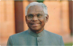
Shri. K. R. Narayanan
Past President Of India
I was delighted to visit Shroff’s Eye Hospital and see that it has been refurbished and expanded in a Spacious and beautiful manner. This well equipped and competently staffed Eye Centre has become a facility of excellence which will be a boon to the people of Delhi. I wish the Shroff Eye Centre the people and saving their eye sight.
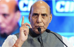
Shri. Raj Nath Singh
Home Minister Of India
I am of firm opinion that Dr. Shroff’s institution is the best eye institution. I am fully satisfied with the care and treatment of my eyes. I had heard lot of praise about this institution, I got the same. I wish all the best for this institution.

Smt. Sheila Dikshit
Chief Minister Of Delhi
This wonderful clinic gives such comfort and confidence. I moved out feeling so confident to see the world.

Smt. Sharmila Tagore
Actor/Director
I was very well looked after, even pampered. The staff is extremely competent. Every single one of them. The whole atmosphere is truly democratic. No one is given more importance than the other. Thank you. Everyone is treated equally.
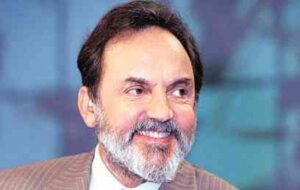
Shri. Prannoy Roy
CEO, NDTV India
This is undoubtedly the finest institution for eyes. Not only is every single staff member highly professional and brilliantly competent, everyone is warm, patient understanding and patient friendly. God bless all of you.

Smt. Shehnaz Hussain
CEO, Shahnaz Herbals
The best eye clinic in the world and Dr. Noshir the last word in eye care.
Media Coverage
Features and Mentions


-
4.9, Based on 10k+ reviews
Our Centres
Eye Hospitals Near You
-
Kailash Colony, South Delhi
A-9, Kailash Colony, New Delhi - 110 048
-
Connaught Place, Central Delhi
105 Surya Kiran, First floor 19, Kasturba Gandhi Marg, New Delhi 110001
-
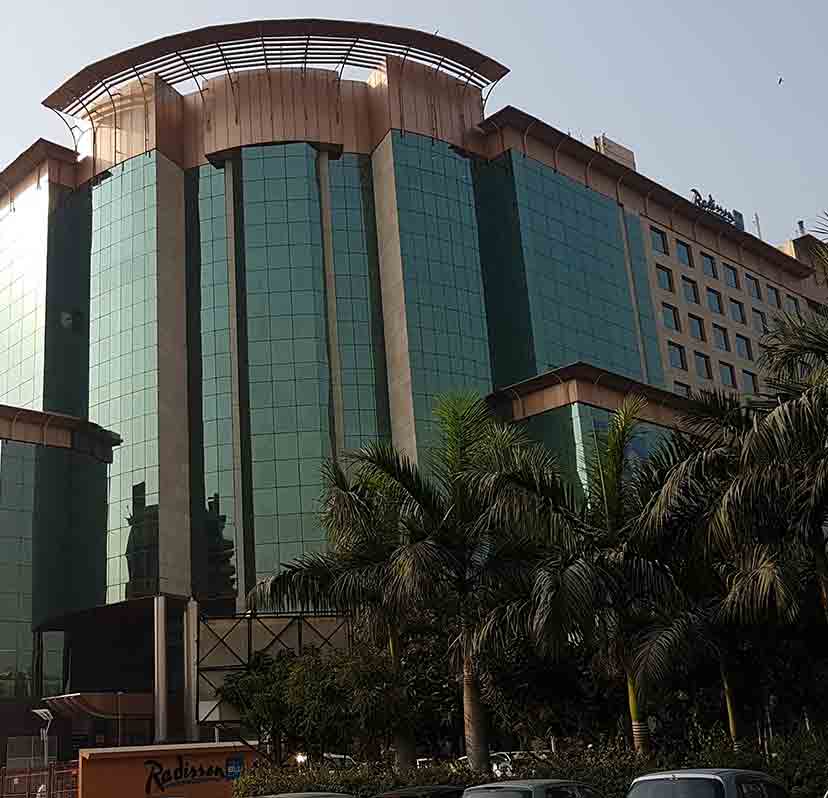
Gurgaon
110, Bestech Chambers (Radisson Suites), B Block, Sushant Lok Phase I, Sector 27, Gurgaon, Haryana - 122002
-
Ghaziabad
509, KM Trade Tower, adjacent to Radisson Blu, Sector 14, Kaushambi, Ghaziabad, Uttar Pradesh 201010